Tuesday, August 14, 2007

Recommendations on uptodate for treatment of Parkinson Disease

SUMMARY AND RECOMMENDATIONS — Either levodopa or a dopamine agonist (DA) can be used initially for patients who require symptomatic therapy [92-94]. Practitioners should always try to find the lowest but still effective dose of dopaminergic medication, either singly or in combination, for patients with PD, each of whom must be evaluated and managed in a highly individual way.
Levodopa (combined with a peripheral decarboxylase inhibitor, ie, Sinemet, Madopar, or Prolopa) is the most effective symptomatic therapy for Parkinson's disease (PD) and should be introduced when the patient and physician jointly decide that quality of life, particularly related to job performance or self care, is substantially compromised. However, levodopa is associated with a higher risk of dyskinesia than the DAs. There does not appear to be a benefit of initiating treatment with controlled release levodopa compared with the immediate release preparation, and the former may limit the ability to follow the initial response to therapy. As a result, it is recommended that therapy be initiated with an immediate release preparation with a subsequent switch to controlled release if indicated. (See "Levodopa" above).
With the exception of pergolide and cabergoline, the dopamine agonists are a useful group of drugs that may be used either as monotherapy in early PD or in combination with other antiparkinsonian drugs for treatment of more advanced disease. They are ineffective in patients who show no response to levodopa. They possibly delay initiation of levodopa therapy and the subsequent appearance of levodopa dyskinesia and motor fluctuations, but at the risk of slightly less efficacy and increased adverse effects [15,51]. (See "Dopamine agonists" above).
Pergolide and cabergoline should not be used for PD because of the risk of valvular heart disease. (See "Valvular heart disease" above).
Selegiline has mild symptomatic benefit, and it may be used in patients with early PD [1,94]. Its use should be limited to patients with early disease since the symptomatic benefits are unlikely to be significant in those with more advanced PD. Nevertheless, patients should understand that there may not be much symptomatic improvement if selegiline is the initial treatment for early PD, and early follow-up and consideration of additional symptomatic therapy should be arranged. The value of selegiline for neuroprotection is unclear [94]. (See "MAO B inhibitors" above).

It is reasonable to initiate therapy with a DA in younger patients (age <65)>65). However, there are exceptions to these general rules, and all treatments should be individualized. Levodopa is the drug of choice if symptoms seriously threaten the patient's lifestyle.

Anticholinergic drugs should be reserved for younger patients in whom tremor is the predominant problem. Their use in older or demented individuals and those without tremor is strongly discouraged.

Sunday, August 12, 2007

MDS and MPS

  • The myelodysplastic syndromes (MDS) are characterized by dysplastic bone marrow hyperplasia or even occasional hypoplasia, associated with variable degrees of peripheral blood cytopenia with or without monocytosis [4]. The paradox between the proliferative bone marrow and the cytopenic peripheral blood picture in the MDS may be explained by increased intramedullary myeloid precursor cell apoptosis [5]. (See "Treatment and prognosis of the myelodysplastic syndromes").
  • The myeloproliferative disorders (MPDs), unlike the MDS, usually exhibit terminal myeloid cell expansion in the peripheral blood [6]. The MPDs are operationally classified into "classic" and "atypical" MPDs. Classic MPDs include polycythemia vera, essential thrombocytosis, chronic myelogenous leukemia (CML), and chronic idiopathic myelofibrosis (AMM, agnogenic myeloid metaplasia).

FAB classification — Patients with MDS have been classified into subgroups based, in part, upon the percentage of bone marrow (BM) blasts, according to a 1982 French, American, and British (FAB) consensus conference (show table 1) [4]:

  • Refractory anemia (RA) — less than 5 percent BM blasts.
  • Refractory anemia with ringed sideroblasts (RARS) — less than 5 percent BM blasts with >15 percent ringed sideroblasts
  • Refractory anemia with excess blasts (RAEB) — 5 to 19 percent BM blasts. RAEB has been further subdivided, with RAEB-I and RAEB-II having BM blast counts of 5-9 and 10-19 percent, respectively [5]
  • Chronic myelomonocytic leukemia (CMML) — up to 20 percent BM blasts plus peripheral blood monocyte count >1000/microL. CMML has been divided into two subtypes: patients with a white blood cell count (WBC) less than or equal to12,000/microL who are considered to have MDS, and the proliferative-type CMML with a WBC >12,000/microL, who are considered to have a chronic myeloproliferative disorder [5]. Based on retrospective data, a separate prognostic scoring system for CMML has been proposed [6]. Unlike the other categories of MDS, an absolute lymphocyte count >2,500/microL was found to be an adverse prognostic factor in CMML [6,7].
  • Refractory anemia with excess blasts in transformation (RAEB-T) — 21 to 30 percent BM blasts

Using the FAB classification system and a meta-analysis of a large number of available studies, patients with RAEB and RAEB-T were shown to have relatively poor prognoses, with median survivals ranging from 5 to 12 months (show table 2). In contrast, those with RA and RARS had median survivals of approximately three to six years.

The proportion of individuals who transformed to AML varied similarly, being 40 to 50 percent for patients with RAEB and RAEB-T, compared to 5 to 15 percent in patients with RA and RARS (show table 2). In a separate study evaluating the time to disease evolution, the incidence of transformation to AML at one and two years was zero in RARS, 5 and 10 percent in RA, 25 and 35 percent in RAEB, and 55 and 65 percent in RAEB-T [8].

Saturday, August 11, 2007

Osler Weber Rendu


Pic 1
Osler Weber Rendu syndrome (also known as hereditary hemorrhagic telangiectasia). Note the multiple 1-2 mm, discrete, red macular and papular telangiectases on the lower lip and tongue.

Pic 2
Superficial telangectasias are seen on the lips of a man with hereditary hemorrhagic telangectasia syndrome who had a solitary pulmonary arteriovenous malformation.

Friday, August 10, 2007


Upper panel: bone marrow aspirate stained with Prussian blue to show red cell precursors with numerous iron- positive granules surrounding the nucleus, in some cases forming a complete ring (arrows). Lower panel: Electron micrograph of a single erythroblast with iron-laden (electron dense deposits) mitochondia (red arrows) clustered around its nucleus.

Peripheral blood smear shows basophilic stippling in several red cells from a patient with lead poisoning. The granules represent ribosomal precipitates. A similar picture can be seen in a number of other conditions including thalassemia, megaloblastic anemia, sickle cell anemia, and sideroblastic anemia.


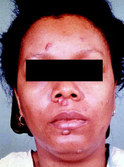
Two types of skin lesions may be seen. The first are the more characteristic verrucous lesions that usually appear on exposed body areas. These often begin as small papulopustular lesions and slowly spread to form crusted, heaped-up lesions that can vary in color from gray to a violaceous hue.


Manifestations of Histoplasmosis
Disseminated histoplasmosis can follow an acute or chronic course. An acute course frequently involves severely immunodepressed patients.
Acute presentations could be as follows-shock, respiratory distress, hepatic, coagulopathy, renal failure, obtundation.
Chronic presentation-The most common presentation is hepatosplenomegaly, elevated liver enzymes, oropharyngeal and gastrointestinal lesions. The skin, CNS, adrenals can be involved also

Manifestations of Coccidoides
Severely immunocompromised
Dyspnea, fever, night sweats, diffuse pulmonary process as in Pneumocystis Carinii

Less immunocompromised
Fever, cough, focal pneumonia

Disseminated coccidoides
Meningitis, skin and lymph node involvement

Manifestations of Blastomycoses
The characteristic rash in blastomycoses-verrucous lesions.


The first are the more characteristic verrucous lesions that usually appear on exposed body areas. These often begin as small papulopustular lesions and slowly spread to form crusted, heaped-up lesions that can vary in color from gray to a violaceous hue.